LECTURE ON THE TOPIC: HUMAN NERVOUS SYSTEM
Nervous system is a system that regulates the activities of all human organs and systems. This system determines: 1) the functional unity of all human organs and systems; 2) connection of the whole organism with environment.
From the point of view of maintaining homeostasis, the nervous system ensures: maintaining the parameters of the internal environment at a given level; inclusion of behavioral responses; adaptation to new conditions if they persist for a long time.
Neuron(nerve cell) - the main structural and functional element of the nervous system; Humans have more than one hundred billion neurons. A neuron consists of a body and processes, usually one long process - an axon and several short branched processes - dendrites. Along dendrites, impulses follow to the cell body, along an axon - from the cell body to other neurons, muscles or glands. Thanks to the processes, neurons contact each other and form neural networks and circles through which nerve impulses circulate.
A neuron is a functional unit of the nervous system. Neurons are susceptible to stimulation, that is, they are capable of being excited and transmitting electrical impulses from receptors to effectors. Based on the direction of impulse transmission, afferent neurons (sensory neurons), efferent neurons (motor neurons) and interneurons are distinguished.
Nervous tissue is called excitable tissue. In response to some impact, a process of excitation arises and spreads in it - rapid recharging of cell membranes. The emergence and propagation of excitation (nerve impulse) is the main way the nervous system carries out its control function.
The main prerequisites for the occurrence of excitation in cells: the existence of an electrical signal on the membrane in a resting state - the resting membrane potential (RMP);
the ability to change the potential by changing the permeability of the membrane for certain ions.
The cell membrane is a semi-permeable biological membrane, it has channels that allow potassium ions to pass through, but there are no channels for intracellular anions, which are retained at the inner surface of the membrane, creating a negative charge of the membrane from the inside; this is the resting membrane potential, which averages - – 70 millivolts (mV). There are 20-50 times more potassium ions in the cell than outside, this is maintained throughout life with the help of membrane pumps (large protein molecules capable of transporting potassium ions from the extracellular environment to the inside). The MPP value is determined by the transfer of potassium ions in two directions:
1. from the outside into the cell under the action of pumps (with a large expenditure of energy);
2. from the cell to the outside by diffusion through membrane channels (without energy consumption).
In the process of excitation, the main role is played by sodium ions, which are always 8-10 times more abundant outside the cell than inside. Sodium channels are closed when the cell is at rest; in order to open them, it is necessary to act on the cell with an adequate stimulus. If the stimulation threshold is reached, the sodium channels open and sodium enters the cell. In thousandths of a second, the membrane charge will first disappear and then change to the opposite - this is the first phase of the action potential (AP) - depolarization. The channels close - the peak of the curve, then the charge is restored on both sides of the membrane (due to potassium channels) - the repolarization stage. The excitation stops and while the cell is at rest, the pumps exchange the sodium that entered the cell for potassium, which left the cell.

An PD evoked at any point on a nerve fiber itself becomes an irritant for neighboring sections of the membrane, causing PD in them, which in turn excite more and more sections of the membrane, thus spreading throughout the entire cell. In fibers covered with myelin, APs will occur only in areas free of myelin. Therefore, the speed of signal propagation increases.


The transfer of excitation from cell to another occurs through a chemical synapse, which is represented by the point of contact of two cells. The synapse is formed by presynaptic and postsynaptic membranes and the synaptic cleft between them. Excitation in the cell resulting from AP reaches the area of the presynaptic membrane where synaptic vesicles are located, from which a special substance, the transmitter, is released. The transmitter entering the gap moves to the postsynaptic membrane and binds to it. Pores open in the membrane for ions, they move into the cell and the process of excitation occurs

Thus, in the cell, the electrical signal is converted into a chemical one, and the chemical signal again into an electrical one. Signal transmission in a synapse occurs more slowly than in a nerve cell, and is also one-sided, since the transmitter is released only through the presynaptic membrane, and can only bind to receptors of the postsynaptic membrane, and not vice versa.
Mediators can cause not only excitation but also inhibition in cells. In this case, pores open on the membrane for ions that strengthen the negative charge that existed on the membrane at rest. One cell can have many synaptic contacts. An example of a mediator between a neuron and a skeletal muscle fiber is acetylcholine.
The nervous system is divided into central nervous system and peripheral nervous system.
In the central nervous system, a distinction is made between the brain, where the main nerve centers and the spinal cord are concentrated, and here there are lower-level centers and pathways to peripheral organs.
Peripheral section - nerves, nerve ganglia, ganglia and plexuses.
The main mechanism of activity of the nervous system is reflex. A reflex is any response of the body to a change in the external or internal environment, which is carried out with the participation of the central nervous system in response to irritation of receptors. The structural basis of the reflex is the reflex arc. It includes five consecutive links:
1 - Receptor - a signaling device that perceives influence;
2 - Afferent neuron – brings a signal from the receptor to the nerve center;
3 - Interneuron – central part of the arc;
4 - Efferent neuron - the signal comes from the central nervous system to the executive structure;
5 - Effector - a muscle or gland performing a certain type of activity

Brain consists of clusters of nerve cell bodies, nerve tracts and blood vessels. Nerve tracts form the white matter of the brain and consist of bundles of nerve fibers that conduct impulses to or from various parts of the gray matter of the brain - nuclei or centers. Pathways connect various nuclei, as well as the brain and spinal cord.
Functionally, the brain can be divided into several sections: the forebrain (consisting of the telencephalon and diencephalon), the midbrain, the hindbrain (consisting of the cerebellum and the pons) and the medulla oblongata. The medulla oblongata, pons, and midbrain are collectively called the brainstem.

Spinal cord located in the spinal canal, reliably protecting it from mechanical damage.
The spinal cord has a segmental structure. Two pairs of anterior and posterior roots extend from each segment, which corresponds to one vertebra. There are 31 pairs of nerves in total.
The dorsal roots are formed by sensory (afferent) neurons, their bodies are located in the ganglia, and the axons enter the spinal cord.
The anterior roots are formed by the axons of efferent (motor) neurons, the bodies of which lie in the spinal cord.
The spinal cord is conventionally divided into four sections - cervical, thoracic, lumbar and sacral. It closes a huge number of reflex arcs, which ensures the regulation of many body functions.
The gray central substance is nerve cells, the white one is nerve fibers.

The nervous system is divided into somatic and autonomic.
TO somatic nervous system (from the Latin word “soma” - body) refers to part of the nervous system (both cell bodies and their processes), which controls the activity of skeletal muscles (body) and sensory organs. This part of the nervous system is largely controlled by our consciousness. That is, we are able to bend or straighten an arm, leg, etc. at will. However, we are unable to consciously stop perceiving, for example, sound signals.
Autonomic nervous system (translated from Latin “vegetative” - plant) is part of the nervous system (both cell bodies and their processes), which controls the processes of metabolism, growth and reproduction of cells, that is, functions common to both animals and plants organisms. The autonomic nervous system is responsible, for example, for the activity of internal organs and blood vessels.
The autonomic nervous system is practically not controlled by consciousness, that is, we are not able to relieve a spasm of the gallbladder at will, stop cell division, stop intestinal activity, dilate or constrict blood vessels
In evolution, the nervous system has undergone several stages of development, which became turning points in the qualitative organization of its activities. These stages differ in the number and types of neuronal formations, synapses, signs of their functional specialization, and the formation of groups of neurons interconnected by common functions. There are three main stages of the structural organization of the nervous system: diffuse, nodular, tubular.
Diffuse The nervous system is the most ancient, found in coelenterates (hydra). Such a nervous system is characterized by a multiplicity of connections between neighboring elements, which allows excitation to freely spread throughout the nervous network in all directions.
This type of nervous system provides wide interchangeability and thereby greater reliability of functioning, but these reactions are imprecise and vague.
Nodal the type of nervous system is typical for worms, mollusks, and crustaceans.

It is characterized by the fact that the connections of nerve cells are organized in a certain way, excitation passes along strictly defined paths. This organization of the nervous system turns out to be more vulnerable. Damage to one node causes dysfunction of the entire organism as a whole, but its qualities are faster and more accurate.
Tubular The nervous system is characteristic of chordates; it includes features of diffuse and nodular types. The nervous system of higher animals took all the best: high reliability of the diffuse type, accuracy, locality, speed of organization of nodal type reactions.
The leading role of the nervous system
At the first stage of the development of the world of living beings, interaction between the simplest organisms was carried out through the aquatic environment of the primitive ocean, into which the chemical substances released by them entered. The first oldest form of interaction between the cells of a multicellular organism is chemical interaction through metabolic products entering the body fluids. Such metabolic products, or metabolites, are the breakdown products of proteins, carbon dioxide, etc. This is the humoral transmission of influences, the humoral mechanism of correlation, or connections between organs.
The humoral connection is characterized by the following features:
- lack of an exact address to which a chemical substance entering the blood or other body fluids is sent;
- the chemical spreads slowly;
- the chemical acts in minute quantities and is usually quickly broken down or eliminated from the body.
Humoral connections are common to both the animal and plant worlds. At a certain stage of development of the animal world, in connection with the appearance of the nervous system, a new, nervous form of connections and regulation is formed, which qualitatively distinguishes the animal world from the plant world. The higher the development of an animal’s organism, the greater the role played by the interaction of organs through the nervous system, which is designated as reflex. In higher living organisms, the nervous system regulates humoral connections. Unlike the humoral connection, the nervous connection has a precise direction to a specific organ and even a group of cells; communication is carried out hundreds of times faster than the speed of distribution of chemicals. The transition from a humoral connection to a nervous connection was not accompanied by the destruction of the humoral connection between the cells of the body, but by the subordination of nervous connections and the emergence of neurohumoral connections.
At the next stage of development of living beings, special organs appear - glands, in which hormones are produced, formed from food substances entering the body. The main function of the nervous system is both to regulate the activity of individual organs among themselves, and in the interaction of the body as a whole with its external environment. Any impact of the external environment on the body appears, first of all, on the receptors (sensory organs) and is carried out through changes caused by the external environment and the nervous system. As the nervous system develops, its highest department—the cerebral hemispheres—becomes “the manager and distributor of all the activities of the body.”
Structure of the nervous system
The nervous system is formed by nervous tissue, which consists of a huge amount neurons- a nerve cell with processes.

The nervous system is conventionally divided into central and peripheral.

Central nervous system includes the brain and spinal cord, and peripheral nervous system- nerves extending from them.

The brain and spinal cord are a collection of neurons. In a cross section of the brain, white and gray matter are distinguished. Gray matter consists of nerve cells, and white matter consists of nerve fibers, which are processes of nerve cells. In different parts of the central nervous system, the location of white and gray matter is different. In the spinal cord, gray matter is located inside, and white matter is outside, but in the brain (cerebral hemispheres, cerebellum), on the contrary, gray matter is outside, white matter is inside. In various parts of the brain there are separate clusters of nerve cells (gray matter) located inside the white matter - kernels. Clusters of nerve cells are also located outside the central nervous system. They are called nodes and belong to the peripheral nervous system.
Reflex activity of the nervous system
The main form of activity of the nervous system is the reflex. Reflex- the body’s reaction to changes in the internal or external environment, carried out with the participation of the central nervous system in response to irritation of receptors.
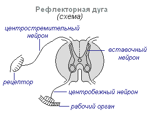
With any irritation, excitation from the receptors is transmitted along centripetal nerve fibers to the central nervous system, from where, through the interneuron along centrifugal fibers, it goes to the periphery to one or another organ, the activity of which changes. This entire path through the central nervous system to the working organ is called reflex arc usually formed by three neurons: sensory, intercalary and motor. A reflex is a complex act in which a significantly larger number of neurons take part. Excitation, entering the central nervous system, spreads to many parts of the spinal cord and reaches the brain. As a result of the interaction of many neurons, the body responds to irritation.
Spinal cord
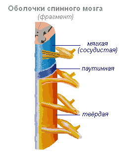
Spinal cord- a cord about 45 cm long, 1 cm in diameter, located in the spinal canal, covered with three meninges: dura, arachnoid and soft (vascular).
Spinal cord is located in the spinal canal and is a cord that at the top passes into the medulla oblongata and at the bottom ends at the level of the second lumbar vertebra. The spinal cord consists of gray matter containing nerve cells and white matter consisting of nerve fibers. Gray matter is located inside the spinal cord and is surrounded on all sides by white matter.

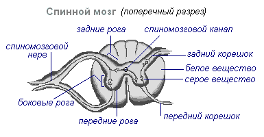
In a cross section, the gray matter resembles the letter H. It distinguishes the anterior and posterior horns, as well as the connecting crossbar, in the center of which there is a narrow canal of the spinal cord containing cerebrospinal fluid. In the thoracic region there are lateral horns. They contain the bodies of neurons that innervate internal organs. The white matter of the spinal cord is formed by nerve processes. Short processes connect sections of the spinal cord, and long ones make up the conductive apparatus of bilateral connections with the brain.
The spinal cord has two thickenings - cervical and lumbar, from which nerves extend to the upper and lower extremities. 31 pairs of spinal nerves arise from the spinal cord. Each nerve begins from the spinal cord with two roots - anterior and posterior. Posterior roots - sensitive consist of processes of centripetal neurons. Their bodies are located in the spinal ganglia. Anterior roots - motor- are processes of centrifugal neurons located in the gray matter of the spinal cord. As a result of the fusion of the anterior and posterior roots, a mixed spinal nerve is formed. The spinal cord contains centers that regulate the simplest reflex acts. The main functions of the spinal cord are reflex activity and conduction of excitation.

The human spinal cord contains reflex centers of the muscles of the upper and lower limbs, sweating and urination. The function of excitation is that impulses from the brain to all areas of the body and back pass through the spinal cord. Centrifugal impulses from organs (skin, muscles) are transmitted through ascending pathways to the brain. Along descending pathways, centrifugal impulses are transmitted from the brain to the spinal cord, then to the periphery, to the organs. When the pathways are damaged, there is a loss of sensitivity in various parts of the body, a violation of voluntary muscle contractions and the ability to move.
Evolution of the vertebrate brain
The formation of the central nervous system in the form of a neural tube first appears in chordates. U lower chordates the neural tube persists throughout life, higher- vertebrates - in the embryonic stage, a neural plate is formed on the dorsal side, which sinks under the skin and curls up into a tube. In the embryonic stage of development, the neural tube forms three swellings in the anterior part - three brain vesicles, from which parts of the brain develop: the anterior vesicle gives the forebrain and diencephalon, the middle vesicle turns into the midbrain, the posterior vesicle forms the cerebellum and medulla oblongata. These five brain regions are characteristic of all vertebrates.
For lower vertebrates- fish and amphibians - characterized by a predominance of the midbrain over other parts. U amphibians The forebrain enlarges somewhat and a thin layer of nerve cells forms in the roof of the hemispheres - the primary medullary vault, the ancient cortex. U reptiles The forebrain increases significantly due to accumulations of nerve cells. Most of the roof of the hemispheres is occupied by the ancient cortex. For the first time in reptiles, the rudiment of a new cortex appears. The hemispheres of the forebrain creep onto other parts, as a result of which a bend is formed in the region of the diencephalon. Beginning with ancient reptiles, the cerebral hemispheres become the largest part of the brain.
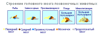
In the structure of the brain birds and reptiles a lot in common. On the roof of the brain is the primary cortex, the midbrain is well developed. However, in birds, compared to reptiles, the total brain mass and the relative size of the forebrain increase. The cerebellum is large and has a folded structure. U mammals the forebrain reaches its greatest size and complexity. Most of the brain matter is made up of the neocortex, which serves as the center of higher nervous activity. The intermediate and middle parts of the brain in mammals are small. The expanding hemispheres of the forebrain cover them and crush them under themselves. Some mammals have a smooth brain without grooves or convolutions, but most mammals have grooves and convolutions in the cerebral cortex. The appearance of grooves and convolutions occurs due to the growth of the brain with limited dimensions of the skull. Further growth of the cortex leads to the appearance of folding in the form of grooves and convolutions.
Brain
If the spinal cord in all vertebrates is developed more or less equally, then the brain differs significantly in size and complexity of structure in different animals. The forebrain undergoes particularly dramatic changes during evolution. In lower vertebrates, the forebrain is poorly developed. In fish, it is represented by the olfactory lobes and nuclei of gray matter in the thickness of the brain. The intensive development of the forebrain is associated with the emergence of animals onto land. It differentiates into the diencephalon and two symmetrical hemispheres, which are called telencephalon. Gray matter on the surface of the forebrain (cortex) first appears in reptiles, developing further in birds and especially in mammals. Truly large forebrain hemispheres become only in birds and mammals. In the latter, they cover almost all other parts of the brain.
The brain is located in the cranial cavity. It includes the brainstem and telencephalon (cerebral cortex).

Brain stem consists of the medulla oblongata, pons, midbrain and diencephalon.
Medulla oblongata is a direct continuation of the spinal cord and, expanding, passes into the hindbrain. It basically retains the shape and structure of the spinal cord. In the thickness of the medulla oblongata there are accumulations of gray matter - the nuclei of the cranial nerves. The rear axle includes cerebellum and pons. The cerebellum is located above the medulla oblongata and has a complex structure. On the surface of the cerebellar hemispheres, gray matter forms the cortex, and inside the cerebellum - its nuclei. Like the spinal medulla oblongata, it performs two functions: reflex and conductive. However, the reflexes of the medulla oblongata are more complex. This is reflected in its importance in the regulation of cardiac activity, the condition of blood vessels, respiration, and sweating. The centers of all these functions are located in the medulla oblongata. Here are the centers for chewing, sucking, swallowing, saliva and gastric juice. Despite its small size (2.5–3 cm), the medulla oblongata is a vital part of the central nervous system. Damage to it can cause death due to cessation of breathing and heart activity. The conductor function of the medulla oblongata and the pons is to transmit impulses from the spinal cord to the brain and back.
IN midbrain the primary (subcortical) centers of vision and hearing are located, which carry out reflexive orienting reactions to light and sound stimulation. These reactions are expressed in various movements of the torso, head and eyes towards the stimuli. The midbrain consists of the cerebral peduncles and quadrigeminalis. The midbrain regulates and distributes the tone (tension) of skeletal muscles.
Diencephalon consists of two departments - thalamus and hypothalamus, each of which consists of a large number of nuclei of the visual thalamus and subthalamic region. Through the visual thalamus, centripetal impulses are transmitted to the cerebral cortex from all receptors of the body. Not a single centripetal impulse, no matter where it comes from, can pass to the cortex, bypassing the visual hillocks. Thus, through the diencephalon, all receptors communicate with the cerebral cortex. In the subtubercular region there are centers that influence metabolism, thermoregulation and endocrine glands.
Cerebellum located behind the medulla oblongata. It consists of gray and white matter. However, unlike the spinal cord and brainstem, the gray matter - the cortex - is located on the surface of the cerebellum, and the white matter is located inside, under the cortex. The cerebellum coordinates movements, makes them clear and smooth, plays an important role in maintaining the balance of the body in space, and also influences muscle tone. When the cerebellum is damaged, a person experiences a decrease in muscle tone, movement disorders and changes in gait, speech slows down, etc. However, after some time, movement and muscle tone are restored due to the fact that the intact parts of the central nervous system take over the functions of the cerebellum.
Large hemispheres- the largest and most developed part of the brain. In humans, they form the bulk of the brain and are covered with cortex over their entire surface. Gray matter covers the outside of the hemispheres and forms the cerebral cortex. The human cerebral cortex has a thickness of 2 to 4 mm and is composed of 6–8 layers formed by 14–16 billion cells, different in shape, size and functions. Under the cortex is a white substance. It consists of nerve fibers connecting the cortex with the lower parts of the central nervous system and the individual lobes of the hemispheres with each other.
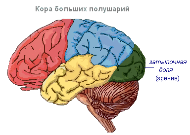
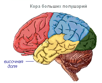
The cerebral cortex has convolutions separated by grooves, which significantly increase its surface. The three deepest grooves divide the hemispheres into lobes. Each hemisphere has four lobes: frontal, parietal, temporal, occipital. Excitation of different receptors enters the corresponding perceptive areas of the cortex, called zones, and from here they are transmitted to a specific organ, prompting it to action. The following zones are distinguished in the cortex. Auditory zone located in the temporal lobe, receives impulses from auditory receptors.

Visual area lies in the occipital region. Impulses from the eye receptors arrive here.
Olfactory zone located on the inner surface of the temporal lobe and is associated with receptors in the nasal cavity.
Sensory-motor the zone is located in the frontal and parietal lobes. This zone contains the main centers of movement of the legs, torso, arms, neck, tongue and lips. This is also where the center of speech lies.

The cerebral hemispheres are the highest division of the central nervous system, controlling the functioning of all organs in mammals. The importance of the cerebral hemispheres in humans also lies in the fact that they represent the material basis of mental activity. I.P. Pavlov showed that mental activity is based on physiological processes occurring in the cerebral cortex. Thinking is associated with the activity of the entire cerebral cortex, and not just with the function of its individual areas.
| Brain department | Functions | |
| Medulla oblongata | Conductor | Connection between the spinal and overlying parts of the brain. |
| Reflex | Regulation of the activity of the respiratory, cardiovascular, digestive systems:
|
|
| Pons | Conductor | Connects the cerebellar hemispheres to each other and to the cerebral cortex. |
| Cerebellum | Coordination | Coordination of voluntary movements and maintaining body position in space. Regulation of muscle tone and balance |
| Midbrain | Conductor | Approximate reflexes to visual and sound stimuli ( turns the head and body). |
| Reflex |
|
|
| Diencephalon | thalamus
hypothalamus
|
|
Cerebral cortex
Surface cerebral cortex in humans is about 1500 cm 2, which is many times greater than inner surface skulls This large surface of the cortex was formed due to the development of a large number of grooves and convolutions, as a result of which most of the cortex (about 70%) is concentrated in the grooves. The largest grooves of the cerebral hemispheres are central, which runs across both hemispheres, and temporal, separating the temporal lobe from the rest. The cerebral cortex, despite its small thickness (1.5–3 mm), has a very complex structure. It has six main layers, which differ in the structure, shape and size of neurons and connections. The cortex contains the centers of all sensory (receptor) systems, representatives of all organs and parts of the body. In this regard, centripetal nerve impulses from all internal organs or parts of the body approach the cortex, and it can control their work. Through the cerebral cortex, conditioned reflexes are closed, through which the body constantly, throughout life, very accurately adapts to the changing conditions of existence, to the environment.
3.1. Origin and functions of the nervous system.
The nervous system in all animals is of ectodermal origin. It performs the following functions:
Communication of the organism with the environment (perception, transmission of irritation and response to irritation);
The connection of all organs and organ systems into a single whole;
The nervous system underlies the formation of higher nervous activity.
3.2. Evolution of the nervous system among invertebrate animals.
The nervous system first appeared in coelenterates and had diffuse or reticular type nervous system, i.e. The nervous system is a network of nerve cells distributed throughout the body and interconnected by thin processes. It has a typical structure in the hydra, but already in jellyfish and polyps clusters of nerve cells appear in certain places(near the mouth, along the edges of the umbrella), these clusters of nerve cells are the precursors of the sensory organs.
Further, the evolution of the nervous system follows the path of concentration of nerve cells in certain places of the body, i.e. along the path of formation of nerve nodes (ganglia). These nodes primarily arise where cells that perceive irritation from the environment are located. Thus, with radial symmetry, a radial type of nervous system arises, and with bilateral symmetry, the concentration of nerve ganglia occurs at the anterior end of the body. Paired nerve trunks extending along the body extend from the head nodes. This type of nervous system is called ganglionic-stem.
This type of nervous system has a typical structure in flatworms, i.e. at the anterior end of the body there are paired ganglia, from which nerve fibers and sensory organs extend forward, and nerve trunks running along the body.
U roundworms the cephalic ganglia merge into the peripharyngeal nerve ring, from which nerve trunks also extend along the body.
In annelids, a nerve chain is formed, i.e. Independent paired nerve nodes are formed in each segment. All of them are connected by both longitudinal and transverse strands. As a result, the nervous system acquires a ladder-like structure. Often both chains come closer together, connecting along the middle part of the body into an unpaired abdominal nerve chain.
Arthropods have the same type of nervous systems, but the number of nerve ganglia decreases and their size increases, especially in the head or cephalothorax, i.e. the process of cephalization is underway.
In mollusks, the nervous system is represented by nodes in different parts of the body, connected to each other by cords and nerves extending from the nodes. Gastropods have pedal, cerebral and pleural-visceral nodes; in bivalves – pedal and pleural-visceral; in cephalopods - pleural-visceral and cerebral nerve ganglia. An accumulation of nervous tissue is observed around the pharynx of cephalopods.
3.3. Evolution of the nervous system in chordates.
The nervous system of chordates is represented by the neural tube, which differentiates into the brain and spinal cord.
In lower chordates, the neural tube has the appearance of a hollow tube (neurocoel) with nerves extending from the tube. In the lancelet, a small expansion is formed in the head section - the rudiment of the brain. This expansion is called the ventricle.
In higher chordates, three swellings are formed at the anterior end of the neural tube: anterior, middle and posterior vesicles. From the first cerebral vesicle, the forebrain and diencephalon are subsequently formed, from the middle cerebral vesicle - the mesencephalon, from the posterior - the cerebellum and medulla oblongata, which passes into the spinal cord.
In all classes of vertebrate animals, the brain consists of 5 sections (anterior, intermediate, middle, posterior and medulla), but the degree of their development is not the same in animals of different classes.
Thus, in cyclostomes, all parts of the brain are located one after another in a horizontal plane. The medulla oblongata directly passes into the spinal cord with the central canal in the nutria.
In fish, the brain is more differentiated compared to cyclostomes. The volume of the forebrain is increased, especially in lungfishes, but the forebrain is not yet divided into hemispheres and functionally serves as the highest olfactory center. The roof of the forebrain is thin, it consists only of epithelial cells and does not contain nerve tissue. In the diencephalon, with which the pineal and pituitary glands are connected, the hypothalamus is located, which is the center of the endocrine system. The most developed in fish is the midbrain. The optic lobes are well expressed in it. In the region of the midbrain there is a bend characteristic of all higher vertebrates. In addition, the midbrain is an analyzing center. The cerebellum, which is part of the hindbrain, is well developed due to the complexity of movement in fish. It represents the center of coordination of movement, its size varies depending on the activity of movement different types fish The medulla oblongata provides communication between the higher parts of the brain and the spinal cord and contains the centers of respiration and circulation.
10 pairs of cranial nerves emerge from the fish brain.
This type of brain, in which the highest center of integration is the midbrain, is called ichthyopsid.
In amphibians, the nervous system in its structure is close to the nervous system of lungfishes, but is distinguished by significant development and complete separation of paired elongated hemispheres, as well as weak development of the cerebellum, which is due to the low mobility of amphibians and the monotony of their movements. But amphibians developed a roof for the forebrain, called the primary medullary vault - archipallium. The number of cranial nerves, like in fish, is ten. And the type of brain is the same, i.e. ichthyopsid.
Thus, all anamnia (cyclostomes, fish and amphibians) have an ichthyopsid type of brain.
In the structure of the brain of reptiles belonging to higher vertebrates, i.e. to amniotes, the features of a progressive organization are clearly expressed. The forebrain hemispheres have a significant predominance over other parts of the brain. At their base there are large accumulations of nerve cells - striatum. Islands of the old cortex, the archicortex, appear on the lateral and medial sides of each hemisphere. The size of the midbrain is reduced, and it loses its importance as a leading center. The bottom of the forebrain becomes the analyzing center, i.e. striped bodies. This type of brain is called sauropsid or striatal. The cerebellum is increased in size due to the variety of movements of reptiles. The medulla oblongata forms a sharp bend, characteristic of all amniotes. There are 12 pairs of cranial nerves leaving the brain.
The same type of brain is characteristic of birds, but with some features. The forebrain hemispheres are relatively large. The olfactory lobes in birds are poorly developed, which indicates the role of smell in the life of birds. In contrast, the midbrain is represented by large optic lobes. The cerebellum is well developed, 12 pairs of nerves emerge from the brain.
The brain in mammals reaches its maximum development. The hemispheres are so large that they cover the midbrain and cerebellum. The cerebral cortex is especially developed, its area is increased due to convolutions and grooves. The cortex has a very complex structure and is called the new cortex - neocortex. A secondary medullary vault, the neopallium, appears. Large olfactory lobes are located in front of the hemispheres. The diencephalon, like other classes, includes the pineal gland, pituitary gland and hypothalamus. The midbrain is relatively small, it consists of four tubercles - the quadrigeminal. The anterior cortex is connected with the visual analyzer, the posterior one with the auditory one. Along with the forebrain, the cerebellum progresses greatly. There are 12 pairs of cranial nerves leaving the brain. The analyzing center is the cerebral cortex. This type of brain is called mammary.
3.4. Anomalies and malformations of the nervous system in humans.
1. Acephaly- absence of the brain, vault, skull and facial skeleton; this disorder is associated with underdevelopment of the anterior neural tube and is combined with defects of the spinal cord, bones and internal organs.
2. Anencephaly- absence of the cerebral hemispheres and skull roof with underdevelopment of the brain stem and is combined with other developmental defects. This pathology is caused by non-closure (dysraphism) of the head of the neural tube. In this case, the bones of the roof of the skull do not develop, and the bones of the base of the skull show various anomalies. Anencephaly is incompatible with life, the average frequency is 1/1500, and is more common in female fetuses.
3. Atelencephaly– arrest of development (heterochrony) of the anterior part of the neural tube at the stage of three vesicles. As a result, the cerebral hemispheres and subcortical nuclei are not formed.
4. Prosencephaly– the telencephalon is divided by a longitudinal groove, but in depth both hemispheres remain connected to each other.
5. Holoprosencephaly– the telencephalon is not divided into hemispheres and has the appearance of a hemisphere with a single cavity (ventricle).
6. Alobar prosencephaly– the division of the telencephalon is only in the posterior part, and the frontal lobes remain undivided.
7. Aplasia or hypoplasia of the corpus callosum– complete or partial absence of a complex commissure of the brain, i.e. corpus callosum.
8. Hydroencephaly- atrophy of the cerebral hemispheres in combination with hydrocephalus.
9. Agiriya- complete absence of grooves and convolutions (smooth brain) of the cerebral hemispheres.
10. Microgyria- reduction in the number and volume of furrows.
11. Congenital hydrocephalus- obstruction of part of the ventricular system of the brain and its outputs, it is caused by a primary disorder of the development of the nervous system.
12. Spina bifida- a defect in the closure and separation of the spinal neural tube from the skin ectoderm. Sometimes this anomaly is accompanied by diplomyelia, in which the spinal cord is split along a certain length into two parts, each with its own central recess.
13. Iniencephaly- a rare anomaly, incompatible with life, occurs more often in female fetuses. This is a gross anomaly of the back of the head and brain. The head is turned so that the face is facing upward. Dorsally, the scalp continues into the skin of the lumbodorsal or sacral region.
In the human body, the work of all its organs is closely interconnected, and therefore the body functions as a single whole. The coordination of the functions of internal organs is ensured by the nervous system, which, in addition, communicates the body as a whole with the external environment and controls the functioning of each organ.
Distinguish central nervous system (brain and spinal cord) and peripheral, represented by nerves extending from the brain and spinal cord and other elements lying outside the spinal cord and brain. The entire nervous system is divided into somatic and autonomic (or autonomic). Somatic nervous the system primarily communicates the body with the external environment: perception of irritations, regulation of movements of the striated muscles of the skeleton, etc., vegetative - regulates metabolism and the functioning of internal organs: heartbeat, peristaltic contractions of the intestines, secretion of various glands, etc. Both of them function in close interaction, but the autonomic nervous system has some independence (autonomy), controlling many involuntary functions.
A cross-section of the brain shows that it consists of gray and white matter. Gray matter is a collection of neurons and their short processes. In the spinal cord it is located in the center, surrounding the spinal canal. In the brain, on the contrary, gray matter is located along its surface, forming a cortex and separate clusters called nuclei, concentrated in the white matter. White matter is located under the gray and is composed of nerve fibers covered with membranes. Nerve fibers, when connected, form nerve bundles, and several such bundles form individual nerves. The nerves through which excitation is transmitted from the central nervous system to the organs are called centrifugal, and the nerves that conduct excitation from the periphery to the central nervous system are called centripetal.
The brain and spinal cord are covered with three membranes: dura mater, arachnoid membrane and vascular membrane. Solid - external, connective tissue, lining the internal cavity of the skull and spinal canal. Arachnoid located under the dura ~ this is a thin shell with a small number of nerves and blood vessels. Vascular the membrane is fused with the brain, extends into the grooves and contains many blood vessels. Between the choroid and arachnoid membranes, cavities filled with brain fluid are formed.
In response to irritation, the nervous tissue enters a state of excitation, which is a nervous process that causes or enhances the activity of the organ. The property of nervous tissue to transmit excitation is called conductivity. The speed of excitation is significant: from 0.5 to 100 m/s, therefore, interaction is quickly established between organs and systems that meets the needs of the body. Excitation is carried out along the nerve fibers in isolation and does not pass from one fiber to another, which is prevented by the membranes covering the nerve fibers.
The activity of the nervous system is reflexive character. The response to stimulation carried out by the nervous system is called reflex. The path along which nervous excitation is perceived and transmitted to the working organ is called reflex arc. It consists of five sections: 1) receptors that perceive irritation; 2) sensitive (centripetal) nerve, transmitting excitation to the center; 3) the nerve center, where excitation switches from sensory neurons to motor neurons; 4) motor (centrifugal) nerve, carrying excitation from the central nervous system to the working organ; 5) a working organ that reacts to the received irritation.
The process of inhibition is the opposite of excitation: it stops activity, weakens or prevents its occurrence. Excitation in some centers of the nervous system is accompanied by inhibition in others: nerve impulses entering the central nervous system can delay certain reflexes. Both processes are excitation And braking - are interconnected, which ensures coordinated activity of organs and the entire organism as a whole. For example, during walking, contraction of the flexor and extensor muscles alternates: when the flexion center is excited, impulses follow to the flexor muscles, at the same time, the extension center is inhibited and does not send impulses to the extensor muscles, as a result of which the latter relax, and vice versa.
Spinal cord is located in the spinal canal and has the appearance of a white cord stretching from the occipital foramen to the lower back. There are longitudinal grooves along the anterior and posterior surfaces of the spinal cord; the spinal canal runs in the center, around which the gray matter - an accumulation of a huge number of nerve cells that form a butterfly outline. Along the outer surface of the spinal cord there is white matter - a cluster of bundles of long processes of nerve cells.
In the gray matter, anterior, posterior and lateral horns are distinguished. They lie in the anterior horns motor neurons, in the rear - insert, which communicate between sensory and motor neurons. Sensory neurons lie outside the cord, in the spinal ganglia along the sensory nerves. Long processes extend from the motor neurons of the anterior horns - anterior roots, forming motor nerve fibers. Axons of sensory neurons approach the dorsal horns, forming back roots, which enter the spinal cord and transmit excitation from the periphery to the spinal cord. Here the excitation switches to the interneuron, and from it to the short processes of the motor neuron, from which it is then communicated to the working organ along the axon.
In the intervertebral foramina, the motor and sensory roots are connected, forming mixed nerves, which then split into front and rear branches. Each of them consists of sensory and motor nerve fibers. Thus, at the level of each vertebra from the spinal cord in both directions only 31 pairs depart mixed type spinal nerves. The white matter of the spinal cord forms pathways that stretch along the spinal cord, connecting both its individual segments with each other and the spinal cord with the brain. Some pathways are called ascending or sensitive, transmitting excitation to the brain, others - downward or motor, which conduct impulses from the brain to certain segments of the spinal cord.
Function of the spinal cord. The spinal cord performs two functions - reflex and conduction.
Each reflex is carried out by a strictly defined part of the central nervous system - the nerve center. A nerve center is a collection of nerve cells located in one of the parts of the brain and regulating the activity of an organ or system. For example, the center of the knee reflex is located in the lumbar spinal cord, the center of urination is in the sacral, and the center of pupil dilation is in the upper thoracic segment of the spinal cord. The vital motor center of the diaphragm is localized in the III-IV cervical segments. Other centers - respiratory, vasomotor - are located in the medulla oblongata. In the future, some more nerve centers that control certain aspects of the body’s life will be considered. The nerve center consists of many interneurons. It processes the information that comes from the corresponding receptors, and generates impulses that are transmitted to the executive organs - the heart, blood vessels, skeletal muscles, glands, etc. As a result of them functional state changes. To regulate the reflex and its accuracy, the participation of the higher parts of the central nervous system, including the cerebral cortex, is necessary.
The nerve centers of the spinal cord are directly connected to the receptors and executive organs of the body. Motor neurons of the spinal cord provide contraction of the muscles of the trunk and limbs, as well as the respiratory muscles - the diaphragm and intercostal muscles. In addition to the motor centers of skeletal muscles, the spinal cord contains a number of autonomic centers.
Another function of the spinal cord is conduction. Bundles of nerve fibers that form white matter connect various parts of the spinal cord to each other and the brain to the spinal cord. There are ascending pathways that carry impulses to the brain, and descending pathways that carry impulses from the brain to the spinal cord. According to the first, excitation arising in the receptors of the skin, muscles, and internal organs is carried along the spinal nerves to the dorsal roots of the spinal cord, perceived by sensitive neurons of the spinal nodes and from here sent either to the dorsal horns of the spinal cord, or as part of the white matter reaches the trunk, and then the cerebral cortex. Descending pathways carry excitation from the brain to the motor neurons of the spinal cord. From here, excitation is transmitted along the spinal nerves to the executive organs.
The activity of the spinal cord is controlled by the brain, which regulates spinal reflexes.
Brain located in the brain part of the skull. Its average weight is 1300-1400 g. After a person is born, brain growth continues up to 20 years. It consists of five sections: the anterior (cerebral hemispheres), intermediate, middle "hindbrain and medulla oblongata. Inside the brain there are four interconnected cavities - cerebral ventricles. They are filled with cerebrospinal fluid. The first and second ventricles are located in the cerebral hemispheres, the third - in the diencephalon, and the fourth - in the medulla oblongata. The hemispheres (the newest part in evolutionary terms) reach in humans high development, making up 80% of the brain mass. The phylogenetically more ancient part is the brain stem. The trunk includes the medulla oblongata, pons, midbrain and diencephalon. The white matter of the trunk contains numerous nuclei of gray matter. The nuclei of 12 pairs of cranial nerves also lie in the brain stem. The brainstem is covered by the cerebral hemispheres.
The medulla oblongata is a continuation of the spinal cord and repeats its structure: there are also grooves on the anterior and posterior surfaces. It consists of white matter (conducting bundles), where clusters of gray matter are scattered - the nuclei from which cranial nerves originate - from the IX to the XII pairs, including the glossopharyngeal (IX pair), vagus (X pair), innervating the respiratory organs, blood circulation, digestion and other systems, sublingual (XII pair).. At the top, the medulla oblongata continues into a thickening - pons, and from the sides why the lower cerebellar peduncles extend. From above and from the sides, almost the entire medulla oblongata is covered by the cerebral hemispheres and the cerebellum.
The gray matter of the medulla oblongata contains vital centers that regulate cardiac activity, breathing, swallowing, carrying out protective reflexes (sneezing, coughing, vomiting, lacrimation), secretion of saliva, gastric and pancreatic juice, etc. Damage to the medulla oblongata can cause death due to the cessation of cardiac activity and respiration.
The hindbrain includes the pons and cerebellum. Pons It is bounded below by the medulla oblongata, from above it passes into the cerebral peduncles, and its lateral sections form the middle peduncles of the cerebellum. The substance of the pons contains the nuclei of the V to VIII pairs of cranial nerves (trigeminal, abducens, facial, auditory).
Cerebellum located posterior to the pons and medulla oblongata. Its surface consists of gray matter (cortex). Under the cerebellar cortex there is white matter, in which there are accumulations of gray matter - the nuclei. The entire cerebellum is represented by two hemispheres, the middle part - the vermis and three pairs of legs formed by nerve fibers, through which it is connected to other parts of the brain. The main function of the cerebellum is unconditioned reflex coordination of movements, which determines their clarity, smoothness and preservation of body balance, as well as maintaining muscle tone. Through the spinal cord, along the pathways, impulses from the cerebellum enter the muscles.
The cerebral cortex controls the activity of the cerebellum. The midbrain is located in front of the pons and is represented by quadrigeminal And legs of the brain. In its center there is a narrow canal (brain aqueduct), which connects the III and IV ventricles. The cerebral aqueduct is surrounded by gray matter, in which the nuclei of the III and IV pairs of cranial nerves lie. In the cerebral peduncles the pathways from the medulla oblongata continue; pons to the cerebral hemispheres. The midbrain plays an important role in the regulation of tone and in the implementation of reflexes that make standing and walking possible. The sensitive nuclei of the midbrain are located in the quadrigeminal tubercles: the upper ones contain nuclei associated with the organs of vision, and the lower ones contain nuclei associated with the organs of hearing. With their participation, orienting reflexes to light and sound are carried out.
The diencephalon occupies the highest position in the brainstem and lies anterior to the cerebral peduncles. Consists of two visual tuberosities, supracubertal, subtubercular region and geniculate bodies. Along the periphery of the diencephalon there is white matter, and in its thickness there are nuclei of gray matter. Visual tuberosities - the main subcortical centers of sensitivity: impulses from all receptors of the body arrive here along the ascending pathways, and from here to the cerebral cortex. In the sub-hillock part (hypothalamus) there are centers, the totality of which represents the highest subcortical center of the autonomic nervous system, regulating metabolism in the body, heat transfer, and the constancy of the internal environment. The parasympathetic centers are located in the anterior parts of the hypothalamus, and the sympathetic centers in the posterior parts. The subcortical visual and auditory centers are concentrated in the nuclei of the geniculate bodies.
The second pair of cranial nerves, the optic ones, goes to the geniculate bodies. The brain stem is connected to the environment and to the organs of the body by cranial nerves. By their nature they can be sensitive (I, II, VIII pairs), motor (III, IV, VI, XI, XII pairs) and mixed (V, VII, IX, X pairs).
Autonomic nervous system. Centrifugal nerve fibers are divided into somatic and autonomic. Somatic conduct impulses to skeletal striated muscles, causing them to contract. They originate from motor centers located in the brainstem, in the anterior horns of all segments of the spinal cord and, without interruption, reach the executive organs. Centrifugal nerve fibers going to internal organs and systems, to all tissues of the body, are called vegetative. Centrifugal neurons of the autonomic nervous system lie outside the brain and spinal cord - in the peripheral nerve nodes - ganglia. The processes of ganglion cells end in smooth muscle, cardiac muscle and glands.
The function of the autonomic nervous system is to regulate physiological processes in the body, to ensure the body's adaptation to changing environmental conditions.
The autonomic nervous system does not have its own special sensory pathways. Sensitive impulses from organs are sent along sensory fibers common to the somatic and autonomic nervous systems. The regulation of the autonomic nervous system is carried out by the cerebral cortex.
The autonomic nervous system consists of two parts: sympathetic and parasympathetic. Nuclei of the sympathetic nervous system located in the lateral horns of the spinal cord, from the 1st thoracic to the 3rd lumbar segments. Sympathetic fibers leave the spinal cord as part of the anterior roots and then enter the nodes, which, connected by short bundles in a chain, form a paired border trunk located on both sides of the spinal column. Next, from these nodes, the nerves go to the organs, forming plexuses. Impulses entering the organs through sympathetic fibers provide reflex regulation of their activity. They strengthen and increase heart rate, cause rapid redistribution of blood by narrowing some vessels and dilating others.
Parasympathetic nerve nuclei lie in the middle, medulla oblongata and sacral parts of the spinal cord. Unlike the sympathetic nervous system, all parasympathetic nerves reach peripheral nerve nodes located in the internal organs or on the approaches to them. The impulses conducted by these nerves cause a weakening and slowing of cardiac activity, a narrowing of the coronary vessels of the heart and brain vessels, dilation of the vessels of the salivary and other digestive glands, which stimulates the secretion of these glands, and increases the contraction of the muscles of the stomach and intestines.
Most internal organs receive dual autonomic innervation, that is, they are approached by both sympathetic and parasympathetic nerve fibers, which function in close interaction, exerting the opposite effect on the organs. It has great value in adapting the body to constantly changing environmental conditions.
The forebrain consists of highly developed hemispheres and the middle part connecting them. The right and left hemispheres are separated from each other by a deep fissure at the bottom of which lies the corpus callosum. Corpus callosum connects both hemispheres through long processes of neurons that form pathways. The cavities of the hemispheres are represented lateral ventricles(I and II). The surface of the hemispheres is formed by gray matter or the cerebral cortex, represented by neurons and their processes; under the cortex lies white matter - pathways. Pathways connect individual centers within one hemisphere, or the right and left halves of the brain and spinal cord, or different floors of the central nervous system. The white matter also contains clusters of nerve cells that form the subcortical nuclei of the gray matter. Part of the cerebral hemispheres is the olfactory brain with a pair of olfactory nerves extending from it (I pair).
The total surface of the cerebral cortex is 2000 - 2500 cm 2, its thickness is 2.5 - 3 mm. The cortex includes more than 14 billion nerve cells arranged in six layers. In a three-month-old embryo, the surface of the hemispheres is smooth, but the cortex grows faster than the braincase, so the cortex forms folds - convolutions, limited by grooves; they contain about 70% of the surface of the cortex. Furrows divide the surface of the hemispheres into lobes. Each hemisphere has four lobes: frontal, parietal, temporal And occipital, The deepest grooves are the central ones, separating the frontal lobes from the parietal lobes, and the lateral ones, which delimit the temporal lobes from the rest; The parieto-occipital sulcus separates the parietal lobe from the occipital lobe (Fig. 85). Anterior to the central sulcus in the frontal lobe is the anterior central gyrus, behind it is the posterior central gyrus. The lower surface of the hemispheres and the brain stem is called base of the brain.
To understand how the cerebral cortex functions, you need to remember that the human body has large number a variety of highly specialized receptors. Receptors are capable of detecting the most minor changes in the external and internal environment.
Receptors located in the skin respond to changes in the external environment. In muscles and tendons there are receptors that signal to the brain about the degree of muscle tension and joint movements. There are receptors that respond to changes in the chemical and gas composition of the blood, osmotic pressure, temperature, etc. In the receptor, irritation is converted into nerve impulses. Along sensitive nerve pathways, impulses are carried to the corresponding sensitive zones of the cerebral cortex, where a specific sensation is formed - visual, olfactory, etc.
The functional system, consisting of a receptor, a sensitive pathway and a zone of the cortex where this type of sensitivity is projected, was called by I. P. Pavlov analyzer.
Analysis and synthesis of the received information is carried out in a strictly defined area - the zone of the cerebral cortex. The most important areas of the cortex are motor, sensitive, visual, auditory, and olfactory. Motor the zone is located in the anterior central gyrus in front of the central sulcus of the frontal lobe, the zone musculocutaneous sensitivity - behind the central sulcus, in the posterior central gyrus of the parietal lobe. Visual the zone is concentrated in the occipital lobe, auditory - in the superior temporal gyrus of the temporal lobe, and olfactory And gustatory zones - in the anterior temporal lobe.
The activity of analyzers reflects the external material world. This enables mammals to adapt to environmental conditions by changing behavior. Man, learning natural phenomena, the laws of nature and creating tools, actively changes the external environment, adapting it to his needs.
Many neural processes take place in the cerebral cortex. Their purpose is twofold: interaction of the body with the external environment (behavioral reactions) and the unification of body functions, nervous regulation of all organs. The activity of the cerebral cortex of humans and higher animals was defined by I. P. Pavlov as higher nervous activity, representing conditioned reflex function cerebral cortex. Even earlier, the main principles about the reflex activity of the brain were expressed by I. M. Sechenov in his work “Reflexes of the Brain.” However modern performance about higher nervous activity was created by I.P. Pavlov, who, by studying conditioned reflexes, substantiated the mechanisms of adaptation of the body to changing environmental conditions.
Conditioned reflexes are developed during the individual life of animals and humans. Therefore, conditioned reflexes are strictly individual: some individuals may have them, while others may not. For such reflexes to occur, the action of the conditioned stimulus must coincide in time with the action of the unconditioned stimulus. Only the repeated coincidence of these two stimuli leads to the formation of a temporary connection between the two centers. According to the definition of I.P. Pavlov, reflexes acquired by the body during its life and resulting from the combination of indifferent stimuli with unconditioned ones are called conditioned.
In humans and mammals, new conditioned reflexes are formed throughout life; they are locked in the cerebral cortex and are temporary in nature, since they represent temporary connections of the organism with the environmental conditions in which it is located. Conditioned reflexes in mammals and humans are very complex to develop, since they cover a whole complex of stimuli. In this case, connections arise between different parts of the cortex, between the cortex and subcortical centers, etc. The reflex arc becomes significantly more complex and includes receptors that perceive conditioned stimulation, a sensory nerve and the corresponding pathway with subcortical centers, a section of the cortex that perceives conditioned irritation, second area associated with the center of the unconditioned reflex, center of the unconditioned reflex, motor nerve, working organ.
During the individual life of an animal and a person, countless formed conditioned reflexes serve as the basis for his behavior. Animal training is also based on the development of conditioned reflexes, which arise as a result of combination with unconditioned ones (giving treats or encouraging affection) when jumping through a burning ring, lifting on their paws, etc. Training is important in the transportation of goods (dogs, horses), border protection, hunting (dogs), etc.
Various environmental stimuli acting on the body can cause not only the formation of conditioned reflexes in the cortex, but also their inhibition. If inhibition occurs immediately upon the first action of the stimulus, it is called unconditional. When braking, suppression of one reflex creates conditions for the emergence of another. For example, the smell of a predatory animal inhibits the consumption of food by a herbivore and causes an orienting reflex, in which the animal avoids meeting the predator. In this case, in contrast to unconditional inhibition, the animal develops conditioned inhibition. It occurs in the cerebral cortex when a conditioned reflex is reinforced by an unconditioned stimulus and ensures the animal’s coordinated behavior in constantly changing environmental conditions, when useless or even harmful reactions are excluded.
Higher nervous activity. Human behavior is associated with conditioned-unconditioned reflex activity. Based on unconditioned reflexes, starting from the second month after birth, the child develops conditioned reflexes: as he develops, communicates with people and is influenced by the external environment, temporary connections constantly arise in the cerebral hemispheres between their various centers. The main difference between human higher nervous activity is thinking and speech, which appeared as a result of labor social activity. Thanks to the word, generalized concepts and ideas arise, as well as the ability for logical thinking. As a stimulus, a word evokes a large number of conditioned reflexes in a person. They are the basis for training, education, and the development of work skills and habits.
Based on the development of speech function in people, I. P. Pavlov created the doctrine of first and second signaling systems. The first signaling system exists in both humans and animals. This system, the centers of which are located in the cerebral cortex, perceives through receptors direct, specific stimuli (signals) of the external world - objects or phenomena. In humans, they create the material basis for sensations, ideas, perceptions, impressions of surrounding nature and social environment, and this forms the basis concrete thinking. But only in humans there is a second signaling system associated with the function of speech, with the word audible (speech) and visible (writing).
A person can be distracted from the characteristics of individual objects and find common properties in them, which are generalized in concepts and united by one word or another. For example, the word “birds” summarizes representatives of various genera: swallows, tits, ducks and many others. Likewise, every other word acts as a generalization. For a person, a word is not only a combination of sounds or an image of letters, but first of all a form of representing material phenomena and objects of the surrounding world in concepts and thoughts. With the help of words they are formed general concepts. Through the word, signals about specific stimuli are transmitted, and in this case the word serves as a fundamentally new stimulus - signal signals.
When generalizing various phenomena, a person discovers natural connections between them - laws. A person’s ability to generalize is the essence abstract thinking, which distinguishes him from animals. Thinking is the result of the function of the entire cerebral cortex. The second signaling system arose as a result of a joint labor activity people, in which speech became a means of communication between them. On this basis, verbal human thinking arose and developed further. The human brain is the center of thinking and the center of speech associated with thinking.
The dream and its meaning. According to the teachings of I.P. Pavlov and other domestic scientists, sleep is a deep protective inhibition that prevents overwork and exhaustion of nerve cells. It covers the cerebral hemispheres, midbrain and diencephalon. In
During sleep, the activity of many physiological processes drops sharply; only the parts of the brain stem that regulate vital functions continue to function. important functions, - breathing, heartbeat, but their function is also reduced. The sleep center is located in the hypothalamus of the diencephalon, in the anterior nuclei. The posterior nuclei of the hypothalamus regulate the state of awakening and wakefulness.
Monotonous speech, quiet music, general silence, darkness, and warmth help the body fall asleep. During partial sleep, some “sentinel” points of the cortex remain free from inhibition: the mother sleeps soundly when there is noise, but the slightest rustle of the child wakes her up; soldiers sleep with the roar of guns and even on the march, but immediately respond to the orders of the commander. Sleep reduces the excitability of the nervous system, and therefore restores its functions.
Sleep comes quickly if stimuli that interfere with the development of inhibition, such as loud music, bright lights, etc., are eliminated.
Using a number of techniques, preserving one excited area, it is possible to induce artificial inhibition in the cerebral cortex (dream-like state) in a person. This condition is called hypnosis. I.P. Pavlov considered it as a partial inhibition of the cortex limited to certain zones. With the onset of the deepest phase of inhibition, weak stimuli (for example, a word) are more effective than strong ones (pain), and high suggestibility is observed. This state of selective inhibition of the cortex is used as a therapeutic technique, during which the doctor instills in the patient that it is necessary to eliminate harmful factors - smoking and drinking alcohol. Sometimes hypnosis can be caused by a strong, unusual stimulus under given conditions. This causes “numbness,” temporary immobilization, and concealment.
Dreams. Both the nature of sleep and the essence of dreams are revealed on the basis of the teachings of I.P. Pavlov: during a person’s wakefulness, excitation processes predominate in the brain, and when all areas of the cortex are inhibited, complete deep sleep develops. With such sleep there are no dreams. In the case of incomplete inhibition, individual uninhibited brain cells and areas of the cortex enter into various interactions with each other. Unlike normal connections in the waking state, they are characterized by quirkiness. Every dream is a more or less vivid and complex event, a picture, a living image that periodically arises in a sleeping person as a result of the activity of cells that remain active during sleep. According to I.M. Sechenov, “dreams are unprecedented combinations of experienced impressions.” Often, external irritations are included in the content of a dream: a warmly covered person sees himself in hot countries, the cooling of his feet is perceived by him as walking on the ground, in the snow, etc. Scientific analysis of dreams from a materialistic point of view has shown the complete failure of the predictive interpretation of “prophetic dreams.”
Hygiene of the nervous system. The functions of the nervous system are carried out by balancing excitatory and inhibitory processes: excitation at some points is accompanied by inhibition at others. At the same time, the functionality of the nervous tissue is restored in the areas of inhibition. Fatigue is promoted by low mobility during mental work and monotony during physical work. Fatigue of the nervous system weakens its regulatory function and can provoke the occurrence of a number of diseases: cardiovascular, gastrointestinal, skin, etc.
The most favorable conditions for the normal functioning of the nervous system are created with the correct alternation of work, active rest and sleep. Elimination of physical fatigue and nervous fatigue occurs when switching from one type of activity to another, in which different groups of nerve cells will alternately experience the load. In conditions of high automation of production, the prevention of overwork is achieved by the personal activity of the employee, his creative interest, and the regular alternation of moments of work and rest.
Drinking alcohol and smoking cause great harm to the nervous system.
The human nervous system is a stimulator of the muscular system, which we talked about in. As we already know, muscles are needed to move body parts in space, and we have even studied specifically which muscles are intended for which work. But what powers the muscles? What and how makes them work? This will be discussed in this article, from which you will learn the necessary theoretical minimum for mastering the topic indicated in the title of the article.
First of all, it is worth informing that the nervous system is designed to transmit information and commands to our body. The main functions of the human nervous system are the perception of changes within the body and the space surrounding it, the interpretation of these changes and the response to them in the form of a certain form (including muscle contraction).
Nervous system– many different nerve structures interacting with each other, providing, along with endocrine system coordinated regulation of the work of most of the body's systems, as well as response to changing conditions of the external and internal environment. This system combines sensitization, motor activity and the correct functioning of systems such as endocrine, immune and more.
Structure of the nervous system
Excitability, irritability and conductivity are characterized as functions of time, that is, it is a process that occurs from irritation to the appearance of an organ response. The propagation of a nerve impulse in a nerve fiber occurs due to the transition of local foci of excitation to adjacent inactive areas of the nerve fiber. The human nervous system has the property of transforming and generating energies from the external and internal environment and converting them into a nervous process.
Structure of the human nervous system: 1- brachial plexus; 2- musculocutaneous nerve; 3rd radial nerve; 4- median nerve; 5- iliohypogastric nerve; 6-femoral-genital nerve; 7- locking nerve; 8-ulnar nerve; 9 - common peroneal nerve; 10- deep peroneal nerve; 11- superficial nerve; 12- brain; 13- cerebellum; 14- spinal cord; 15- intercostal nerves; 16- hypochondrium nerve; 17 - lumbar plexus; 18-sacral plexus; 19-femoral nerve; 20 - pudendal nerve; 21-sciatic nerve; 22- muscular branches of the femoral nerves; 23- saphenous nerve; 24 tibial nerve
The nervous system functions as a whole with the senses and is controlled by the brain. The largest part of the latter is called the cerebral hemispheres (in the occipital region of the skull there are two smaller hemispheres of the cerebellum). The brain connects to the spinal cord. The right and left cerebral hemispheres are connected to each other by a compact bundle of nerve fibers called the corpus callosum.
Spinal cord- the main nerve trunk of the body - passes through the canal formed by the foramina of the vertebrae and stretches from the brain to the sacral spine. On each side of the spinal cord, nerves extend symmetrically to different parts of the body. The sense of touch is, in general terms, provided by certain nerve fibers, countless endings of which are located in the skin.
Classification of the nervous system
The so-called types of the human nervous system can be represented as follows. The entire integral system is conditionally formed by: the central nervous system - CNS, which includes the brain and spinal cord, and the peripheral nervous system - PNS, which includes numerous nerves extending from the brain and spinal cord. The skin, joints, ligaments, muscles, internal organs and sensory organs send input signals to the central nervous system via PNS neurons. At the same time, outgoing signals from the central nervous system are sent by the peripheral nervous system to the muscles. As visual material, below, the complete human nervous system (diagram) is presented in a logically structured manner.

Central nervous system- the basis of the human nervous system, which consists of neurons and their processes. The main and characteristic function of the central nervous system is the implementation of reflective reactions of varying degrees of complexity, called reflexes. The lower and middle parts of the central nervous system - the spinal cord, medulla oblongata, midbrain, diencephalon and cerebellum - control the activities of individual organs and systems of the body, realize communication and interaction between them, ensure the integrity of the body and its correct functioning. The highest department of the central nervous system - the cerebral cortex and the nearest subcortical formations - for the most part controls the connection and interaction of the body as an integral structure with the outside world.
Peripheral nervous system- is a conditionally allocated part of the nervous system, which is located outside the brain and spinal cord. Includes the nerves and plexuses of the autonomic nervous system, connecting the central nervous system to the organs of the body. Unlike the central nervous system, the PNS is not protected by bones and can be susceptible to mechanical damage. In turn, the peripheral nervous system itself is divided into somatic and autonomic.
- Somatic nervous system- part of the human nervous system, which is a complex of sensory and motor nerve fibers responsible for excitation of muscles, including skin and joints. It also guides the coordination of body movements and the reception and transmission of external stimuli. This system performs actions that a person controls consciously.
- Autonomic nervous system divided into sympathetic and parasympathetic. The sympathetic nervous system controls the response to danger or stress, and can, among other things, cause an increase in heart rate, increased blood pressure and stimulation of the senses by increasing the level of adrenaline in the blood. The parasympathetic nervous system, in turn, controls the state of rest, and regulates the contraction of the pupils, the slowing of the heart rate, the dilation of blood vessels and the stimulation of the digestive and genitourinary systems.

Above you can see a logically structured diagram showing the parts of the human nervous system, in order corresponding to the above material.
Structure and functions of neurons
 All movements and exercises are controlled by the nervous system. The main structural and functional unit of the nervous system (both central and peripheral) is the neuron. Neurons– these are excitable cells that are capable of generating and transmitting electrical impulses (action potentials).
Structure of a nerve cell: 1- cell body; 2- dendrites; 3- cell nucleus; 4- myelin sheath; 5- axon; 6- axon ending; 7- synaptic thickening
The functional unit of the neuromuscular system is the motor unit, which consists of a motor neuron and the muscle fibers it innervates. Actually, the work of the human nervous system, using the process of muscle innervation as an example, occurs as follows.
The cell membrane of the nerve and muscle fiber is polarized, that is, there is a potential difference across it. The inside of the cell contains a high concentration of potassium ions (K), and the outside contains high concentrations of sodium ions (Na). At rest, the potential difference between the inside and outside of the cell membrane does not produce an electrical charge. This specific value is the resting potential. Due to changes in the external environment of the cell, the potential on its membrane constantly fluctuates, and if it increases and the cell reaches its electrical threshold for excitation, there is a sharp change in the electrical charge of the membrane, and it begins to conduct an action potential along the axon to the innervated muscle. By the way, in large muscle groups, one motor nerve can innervate up to 2-3 thousand muscle fibers.
In the diagram below you can see an example of the path a nerve impulse takes from the moment a stimulus occurs to the receipt of a response to it in each individual system.

Nerves connect to each other through synapses, and to muscles through neuromuscular junctions. Synapse- this is the point of contact between two nerve cells, and - the process of transmitting an electrical impulse from a nerve to a muscle.

Synaptic connection: 1- neural impulse; 2- receiving neuron; 3- axon branch; 4- synaptic plaque; 5- synaptic cleft; 6- neurotransmitter molecules; 7- cellular receptors; 8- dendrite of the receiving neuron; 9- synaptic vesicles

Neuromuscular contact: 1- neuron; 2- nerve fiber; 3- neuromuscular contact; 4- motor neuron; 5- muscle; 6- myofibrils
Thus, as we have already said, the process of physical activity in general and muscle contraction in particular is completely controlled by the nervous system.
Conclusion
Today we learned about the purpose, structure and classification of the human nervous system, as well as how it is related to his motor activity and how it affects the functioning of the whole organism as a whole. Since the nervous system is involved in regulating the activity of all organs and systems of the human body, including, and perhaps primarily, the cardiovascular system, then in the next article in the series about the systems of the human body, we will move on to its consideration.